
What we treat
Retina, macula and diabetic eye care
- Age-related macular degeneration
- Diabetic retinopathy
- Retinal tear
- Retinal detachment
- Intravitreal injections
- Vitrectomy
Retina treatment in Patna, Bihar
The retina is like the film in a camera. It is the third and inner coat of the eye — a light-sensitive layer of tissue. When focused light hits the retina, a picture is created and sent to the brain through the optic nerve, giving us vision.
Sometimes part of the retina tears, pulls away or detaches from the back of the eye. When this happens, that part of the retina cannot gather light, and the result is loss of vision.
Diseases and conditions of the retina
- Age-related macular degeneration
- Diabetic retinopathy
- Retinal tear
- Retinal detachment
Age-related macular degeneration (AMD)
What causes AMD?
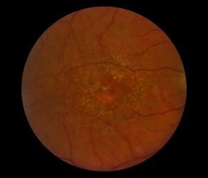
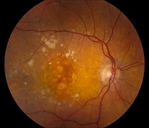
During normal ageing, yellowish deposits called drusen form under the retina, the light-sensitive layer of tissue at the back of the eye that provides clear, sharp images.
As drusen increase in size and number they can interfere with the retina, damaging or killing the light-sensitive cells of the macula. Because these cells give us sharp, detailed vision, the result can be blurring of central vision — and a real impact on reading, driving, or even recognising the face of a friend.
This form is called dry AMD. Dry AMD can be a precursor to wet AMD.
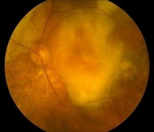
Wet AMD occurs when abnormal blood vessels behind the retina start to grow under the macula. These vessels often leak blood and fluid, damaging light-sensitive cells, and vision is lost quickly. Although about 80 percent of people with AMD have the dry form, wet AMD is responsible for 80 to 90 percent of severe vision loss from the disease.
Living with AMD
AMD can be a difficult condition, but it is not as devastating as people fear when they are diagnosed, and it only rarely leads to complete blindness. Many people live healthy, independent lives with it.
It is important to learn about the disease and work with a retina specialist, who will put a treatment plan in place and can suggest rehabilitation options such as low-vision aids that make everyday living a little easier.
Symptoms
A retina specialist can detect the earliest signs of AMD before you have any vision loss. Those who have had the disease for some time may notice these changes:
Dry AMD
- Need for brighter light when reading
- Difficulty adapting to low light
- Increased blurriness of printed words
- Colours look less bright
- A blurred spot in the centre of vision
- A blank or black spot that starts small and grows over time
Wet AMD
- Sudden, painless decline in central vision
- Straight lines look wavy; objects look larger or smaller than they are
- A well-defined blind spot in the centre of vision
Pay close attention to any decline in central vision, near or distant. If you notice any of these signs, book an examination with a retina specialist.
Risk factors
The main risk factor for AMD is age — the older you are, the greater the risk. People with a family history of AMD are at higher risk, as are women and people of European descent. Some lifestyle factors also raise the risk:
- Cigarette smoking
- Obesity
- High blood pressure
- Excessive sun exposure
- A diet low in fruits and vegetables
Diagnostic tests
Dilated eye exam
Drops widen the pupil so the doctor can examine the retina. If you are over 50, a periodic dilated exam can catch the earliest signs of AMD before vision is lost.
Optical coherence tomography (OCT)
Cross-sectional images of the retina that show its thickness, and whether fluid has leaked into the retinal tissue.
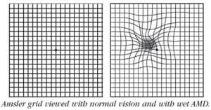
Amsler grid
Cover one eye and look at the dot in the centre. If the lines look broken, wavy or bent, it may be AMD. A normal grid does not rule the disease out.
Visual acuity test
Because AMD affects sharpness of sight, an eye chart can also show that it is developing.
Treatment
There is no known cure for AMD, but early detection and proper treatment can protect vision from further deterioration.
Dry AMD
- Vitamin or mineral supplements to slow its progress
- Keeping to your retina specialist’s examination schedule
- An Amsler grid self-check every two weeks
- Seeing your specialist at once if anything changes
Wet AMD
- Laser surgery — closes abnormal vessels and stops leakage, bleeding or growth
- Photodynamic therapy — a medicine given in the arm is activated by laser to destroy abnormal vessels
- Eye injections — medicines that shut down abnormal vessels and stop them leaking
Intravitreal injections
The anti-VEGF medicines used for wet AMD include:
- Pegaptanib (Macugen)
- Bevacizumab (Avastin)
- Ranibizumab (Lucentis)
Retinal tear and detachment
A tear can result from ageing, an injury to the eye, or another eye problem. A retinal detachment occurs when fluid passes through the tear and separates the retina from the back of the eye.
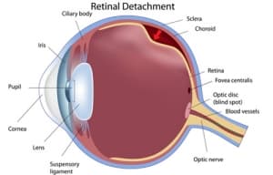
Signs of a retinal tear
- Floaters — specks or threads in your vision
- Flashes — lights, stars or streaks
- Sudden blurry vision
Signs of a retinal detachment
- Flashes of light
- Floaters — small flecks or threads
- Darkening of the side (peripheral) vision
Flashes, a sudden shower of floaters or a shadow over part of your vision need an eye examination the same day.
Sealing a tear — pneumatic retinopexy
Used when the tear is small and easy to close. A small gas bubble is injected into the vitreous gel, where it rises and presses against the retina, closing the tear. Laser or cryopexy then seals it. Each of these treatments fixes the retina to the back of the eye and stops the tear getting worse.
Treating a retinal detachment
Laser treatment of the retina
Reduces leakage, treats abnormal vessel growth, or creates a helpful scar that can prevent a detachment. It is non-invasive.
Scleral buckle
A silicone band is stitched around the white of the eye to push it towards the tear until it heals. The band is not visible and stays in place permanently.
Vitrectomy
A microsurgical technique in which the vitreous gel is removed with a small specialised cutter, under an operating microscope.
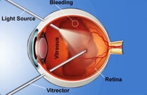
Depending on how complex the detachment is, vitrectomy, buckle, laser and gas bubble may be combined to repair the retina.
Diabetic retinopathy
Diabetic retinopathy is a complication of diabetes that affects the eyes. It is caused by damage to the blood vessels of the retina: when blood sugar stays too high, it harms the small vessels at the back of the eye. It can lead to vision loss or blindness — but there are steps you can take to reduce the risk.
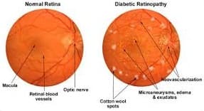
Symptoms
Diabetic retinopathy usually affects both eyes.
- Spots or dark strings floating in your vision (floaters)
- Blurred vision
- Fluctuating vision
- Impaired colour vision
- Dark or empty areas in your vision
- Vision loss
Causes
High blood sugar can damage the small vessels that nourish the retina, and sometimes block them completely, cutting off its blood supply. In response the eye may produce growth factors that lead to diabetic macular oedema, which lowers vision, or proliferative diabetic retinopathy, which can lead to retinal detachment.
Risk factors
- Duration of diabetes — the longer you have had it, the greater the risk
- Poor control of blood sugar
- High blood pressure
- High cholesterol
- Pregnancy
- Tobacco use
Complications
Vitreous haemorrhage
New vessels bleed into the gel that fills the eye. A little blood shows as floaters; a lot can block vision entirely. The blood often clears in weeks or months, and unless the retina is damaged, vision may return.
Retinal detachment
Abnormal vessels stimulate scar tissue that can pull the retina away from the back of the eye, causing floaters, flashes or severe vision loss.
Glaucoma
New vessels growing at the front of the eye can block the fluid’s outflow and raise the eye’s pressure, damaging the optic nerve.
Stages
Background diabetic retinopathy
Damaged retinal vessels leak fluid, protein or fat, bleed, or become blocked.
Proliferative diabetic retinopathy
Blocked vessels starve the retina of oxygen, and fragile new vessels grow that rupture easily, causing large bleeds and severe vision loss.
Diabetic maculopathy
The macula — the centre of the retina — is involved, making near and central vision difficult.
Detection and prevention
There are no symptoms in the early stages, when retinopathy is easiest to treat. By the time vision changes, it is often late — so every person with diabetes needs regular eye check-ups.
- Routine eye examination, as often as your eye doctor advises
- Good control of diabetes
- Control of blood pressure and cholesterol
- Stopping smoking and alcohol
Investigations
Diabetic retinopathy is best diagnosed with a dilated eye exam, in which the doctor looks for abnormal or new vessels, swelling, blood or fatty deposits, scar tissue, bleeding in the vitreous, retinal detachment and changes in the optic nerve. The doctor may also check vision, measure eye pressure and look for cataract.
Fundus photograph
Images of the retina taken with a non-contact fundus camera.
Fundus fluorescein angiography (FFA)
A dye injected into a vein shows, by how it leaks, how severe the retinopathy is and where to treat.
Optical coherence tomography (OCT)
A non-invasive scan — no needle — that with fundus photography gives accurate screening of severity.
Treatment
Laser photocoagulation
The laser spot-welds leaking points in the retina, preventing bleeding, scar tissue and retinal detachment.
Intravitreal injections
A more recent treatment. Not particularly painful, and in many cases more effective than laser alone.
Micro-incision vitrectomy
Keyhole, stitchless surgery that treats complications such as bleeding and retinal detachment.